§1
What we are building
Panacea Bio Chem is developing a four-module system for reversing covalent glycation damage in intact extracellular matrix. The programme is organised around four unknowns rather than around the chemistry, because the chemistry is the part that is already settled.
An engineered flavin-dependent oxidase converts Nε-carboxymethyl-lysine back into native lysine. That reaction works. What decides whether it becomes a repair technology is catalytic rate on protein-bound substrate, transport through dense extracellular matrix, the fate of two reactive byproducts, and whether chemical repair restores any biological function. None of those is resolved, and each is addressed by a hypothesis and a gate below.
Our position
We are not pursuing a faster enzyme first.
The instinct in this area is to attack rate, because rate is the number that improves most readily under directed evolution. We rank access and function above rate, and §4 and §7 give the quantitative argument. A thousandfold faster enzyme that cannot reach its substrate, or whose substrate turns out not to drive the pathology, is a thousandfold faster irrelevance.
The four modules
| # | Module | What it does | Status |
|---|---|---|---|
| 1 | Adduct reversal | Oxidative removal of CML and related side-chain lesions | Lead activity; chemistry demonstrated |
| 2 | Byproduct management | Local disposal of hydrogen peroxide and glyoxylate, measured as a closed balance | Hypothesis 1 |
| 3 | Matrix access | Depth-resolved delivery into intact dermis, soluble first | Hypothesis 3; the binding constraint |
| 4 | Cross-link scission | Selective glucosepane cleavage, enzymatic or chemical | Hypothesis 5; run in parallel from today |
The four are developed independently and combined only once each is separately characterised. They will not share a tissue, a route or a schedule, and treating them as one product is the fastest way to lose the ability to tell which module failed.
§2
The reaction, and the evidence base we start from
The published kinetics set the ceiling on everything downstream, and the ceiling is low: catalytic efficiency is flat at roughly 10 M⁻¹s⁻¹ across every substrate measured. Our programme is designed around that number.
The repair is an oxidative dealkylation. The carboxymethyl group on the lysine ε-amine is oxidised off and the native side chain is released intact:
The catalyst is an engineered FAD-dependent glycine oxidase carrying thirteen substitutions and a two-residue deletion relative to its thermophilic bacterial parent, which was selected for an unusually open active-site channel. Two reactive byproducts leave with every turnover — glyoxylate and hydrogen peroxide — and both are programme concerns in their own right (module 2 and §5).
Published kinetics
Sources for every figure in this section are listed in §16.
| Substrate | Km (mM) | kcat (s⁻¹) | kcat/Km (M⁻¹s⁻¹) | Turnover interval |
|---|---|---|---|---|
| Free CML | 2.2 | 0.040 | 18 | 25 s |
| AA–CML–AA peptide | 0.51 | 0.0053 | 10 | 3.1 min |
| AAA–CML–AAA peptide | 0.14 | 0.0013 | 9.3 | 12.8 min |
Two features of that table drive the whole programme. Affinity improves as the peptide lengthens — Km falls 16-fold — while turnover collapses 31-fold. The enzyme binds extended substrate better and processes it worse. Catalytic efficiency is therefore nearly flat, around 10 M⁻¹s⁻¹: roughly 10–50 times below mature natural protein-editing enzymes and five to six orders of magnitude below diffusion-limited catalysis.
That pattern is not a disappointing number. It is a mechanistic fingerprint, and §6 reads it.
Published tissue performance
| Preparation | Readout | Removal reported |
|---|---|---|
| Aged human arterial sections | Immunohistochemistry | > 70% |
| Aged human skin sections | Immunohistochemistry | > 55% |
| Aged lens protein | Mass spectrometry | 45% |
| Aged lens protein, same preparation | ELISA | 78% |
| Model protein, 33 mapped CML sites | Site-resolved proteomics | 21 sites fell > 50%; 7 fell > 90%; several untouched |
The 45% against 78% on identical lens material is a 33-point gap, and it is the reason no quantitative claim in this programme will rest on an antibody readout. §12 sets out what we use instead.
§3
What that evidence does not establish
None of the published tissue work tests transport, and that is the gap our programme is built to close.
The sections were 4 µm thick, fixed and paraffin-embedded, and incubated with enzyme for 18 hours. In that geometry almost every modified residue sits within a few microns of bulk solvent. Penetration depth is irrelevant by construction. A result obtained there says nothing about 1–3 mm of intact dermis, which is the tissue any therapy has to cross.
Three further limits bear directly on how we design around this evidence.
The site map and the lead enzyme are not the same molecule. The site-resolved proteomics were run on an earlier evolved variant, with an inactive comparator from an earlier generation still. That dataset is evidence for the enzyme family rather than a head-to-head map of the lead variant. Two variants three evolutionary rounds apart behave differently, and we treat the published site map as indicative, not as a specification.
Antibody readouts fail near background. One supplementary value reports 134 ± 61% repair at low initial modification. A removal figure above 100% is a direct demonstration that the readout has stopped measuring anything. Anti-AGE antibodies also recognise CML epitopes in ways that depend on surrounding structure — which is precisely what the enzyme changes — so the readout moves for reasons other than repair.
Partial removal has two explanations and the published work cannot separate them. Removal of 55–70% is consistent with a slow enzyme that ran out of time, and equally consistent with a fast enzyme that finished one pool of sites and never reached another. Those two readings imply opposite development programmes. §7 shows that the published numbers themselves discriminate between them, and that the second reading is the correct one.
We therefore treat the published corpus as establishing one thing only: the chemistry works on accessible substrate. Everything about access, selectivity in real matrix, byproduct fate and biological consequence is open, and is ours to settle.
§4
The seven constraints, ranked
We rank by capacity to stop translation, not by difficulty. Three of the seven can be falsified inside a single well-designed experiment; four are engineering problems with known precedents.
| # | Constraint | Severity | Why it bites | Our resolution |
|---|---|---|---|---|
| 1 | No demonstrated functional benefit | Terminal | Removing a marker is not removing a pathology | Donor-matched viable explants, mechanics and signalling read together |
| 2 | Access to buried sites in intact matrix | Terminal | 4 µm of section is not 4 mm of dermis | Measure reaction depth, not enzyme depth |
| 3 | Turnover on protein-bound CML | High | 12.8 min per catalytic cycle on a hexapeptide | Move the selection pressure onto native matrix |
| 4 | Hydrogen peroxide generation | High | An oxidant produced at the site being repaired | Spatially coupled peroxide disposal |
| 5 | Glyoxylate release | High | Feeds a pathway terminating in oxalate | Full product balance before any dose escalation |
| 6 | Immunogenicity of a bacterial scaffold | High | Repeat dosing against a foreign fold | Epitope depletion, or accept single-site local use |
| 7 | Cross-links untouched | Terminal for mechanics | CML is an adduct, not a bridge | A separate scission chemistry, developed in parallel |
Constraints 1, 2 and 7 decide whether this is a technology at all. Constraints 3 to 6 decide how good a technology it becomes. We resource them in that order.
This ordering is deliberate and it runs against the instinct of the area, which is to attack rate first because rate is the number that improves most readily. Our view is that rate is the cheapest constraint to relieve and the least likely to be the binding one — and that a campaign which relieves it first will have spent its budget before learning whether the target matters.
Constraint 6 deserves one line now, because it looks worse than it is. §13 shows that the natural dosing interval for this therapy is measured in decades, which converts immunogenicity from a recurring-dose liability into a tolerable one.
§5
Hypothesis 1 — redox-coupled repair
Hypothesis. Peroxide disposal co-localised with the repair site, rather than left to bulk tissue defences, will raise net repair per unit of enzyme exposure and reduce collateral oxidation — and the gain will come almost entirely from proximity, not from dose.
The arithmetic is unusually favourable. Catalase turns over up to 2.8 × 10⁶ molecules of peroxide per second. The repair enzyme turns over protein-bound CML at 0.0013 s⁻¹. That is a rate mismatch of roughly 2 × 10⁹.
One catalase molecule can therefore clear the entire peroxide output of two billion repair events per second. Stoichiometric excess is pointless. The only variable that matters is whether the peroxide meets catalase before it meets a susceptible residue — which makes this a geometry problem, not a dosing problem.
That is the ideal coupled net reaction, with catalase returning half the consumed oxygen to the system. In poorly perfused, oxygen-limited matrix this is not cosmetic: the repair reaction is itself oxygen-dependent, and recycling half the oxygen demand raises the depth at which the reaction can still proceed.
Three architectures, in increasing order of commitment
| Architecture | What it buys | What it costs |
|---|---|---|
| Two free enzymes in solution | Simplest; no engineering | Catalase diffuses on its own terms; protection lost wherever the two separate |
| Co-retention in a shared local phase | High local concentration of both, no covalent work | Depends on the phase staying put |
| Fusion or conjugate | Proximity guaranteed | Doubled hydrodynamic radius, directly worsening the transport problem in §7 |
We test in that order, because the third architecture trades constraint 4 against constraint 2, and constraint 2 is the more severe one.
The assay trap
This matters more than the hypothesis.
The standard activity readout for this enzyme class is peroxide production. Adding catalase destroys that readout by design. Any coupled system scored on peroxide fluorescence will appear catalytically dead while working perfectly.
Every coupled experiment in this programme is therefore scored on CML disappearance and lysine appearance by mass spectrometry. This single point invalidates the most obvious experiment anyone would run first, and we expect it to be the most common way a competing group loses a year.
The counter-hypothesis we test in the same experiment
Peroxide at low micromolar concentrations is a physiological signalling species, not only a toxin. Wound-edge peroxide gradients recruit immune cells over 100–200 µm. Measured output in the published protein experiment was about 0.90 µM/hour — squarely in the signalling range, not the damaging one.
It is therefore a live possibility that removing peroxide reduces the biological benefit of repair by suppressing a regenerative signal. Catalase-alone and peroxide-matched arms are not controls here. They are the experiment.
§6
Hypothesis 2 — substrate-realistic evolution
Hypothesis. The enzyme is not rate-limited by chemistry. It is limited by having been selected against substrates that do not resemble its intended target, and the published kinetics carry the fingerprint of that mismatch.
Look again at the three substrates in §2. Km falls from 2.2 mM to 0.14 mM as the peptide lengthens — a 16-fold gain in apparent affinity. Over the same series kcat falls 31-fold. Affinity up, turnover down, efficiency flat.
That pattern has a specific mechanistic meaning: the extended substrate is binding well but binding unproductively. The adduct arrives in the pocket, occupies it, and sits in a geometry from which chemistry cannot proceed. The enzyme is not failing to catch its substrate. It is catching it wrong.
The docking evidence says the same thing. The top-scoring binding pose places the reactive carbon 7.70 Å from the flavin N5 — far outside catalytic range — while a lower-ranked pose sits at 3.69 Å, and dynamics relax the average to 3.58 Å. The best-bound pose is the least productive one. Selecting harder on binding makes this worse, not better.
Why the existing selection system cannot fix it
The directed-evolution scheme that produced this enzyme couples activity to lysine release in a bacterial host. That requires the substrate to be small, soluble and importable. It is an excellent way to find chemistry and a structurally guaranteed way to lose it again at the protein-bound stage. Any screen built on free CML or a hexapeptide is optimising the wrong end of the substrate series.
The quantity to optimise is not kcat. It is the probability that a bound complex is a near-attack conformation:
P(NAC) is the fraction of the bound population within attack distance and angle of the flavin. A tenfold improvement in P(NAC) costs nothing in chemistry and is invisible to an affinity-based screen.
Where we put the mutations
The wider deglycation enzyme family gives an unusually clear structural answer. Fructosamine oxidases share the monomeric sarcosine oxidase fold and have been mapped in detail. Mutational analysis of that family found that altering most active-site residues does little, while the flexible active-site loops are the determinants of what size of substrate the enzyme will accept. That family has sat at exactly this barrier for three decades: excellent on free and low-molecular-weight glycated amines, inert on protein-bound ones.
Our prediction: the residues that matter for protein-bound CML are not in the first shell. They are in the mobile loops and the solvent-facing rim that decide whether a polypeptide backbone can approach at all. Those are precisely the positions a free-substrate screen applies no pressure to.
The tiered selection we substitute
| Tier | Substrate | What it can prove | What it cannot |
|---|---|---|---|
| 1 | Free CML | Chemistry is intact | Anything about proteins |
| 2 | Defined CML peptides | Sequence and steric dependence | Folded-context access |
| 3 | CML on folded globular protein | Attack on a real surface | Matrix architecture |
| 4 | Naturally aged collagen-rich matrix | Authentic accumulated damage | Living-cell response |
| 5 | Viable human tissue explant | Repair in biological context | Systemic behaviour |
Our target is a tenfold gain in repaired sites per unit enzyme exposure measured at tier 4 — not at tier 1, where tenfold is easy and meaningless.
The consequence is worth stating explicitly, because it is a selection rule we will be held to: a variant with a threefold intrinsic gain and markedly better matrix access is the superior molecule, and a kcat-ranked campaign will discard it.
§7
Hypothesis 3 — the penetration–retention paradox
Hypothesis. Tissue repair is not rate-limited. It is limited by a bimodal substrate population — accessible and inaccessible — and by a transport optimum at which stronger matrix binding begins to reduce total repair. Both are calculable from published numbers, and the calculation contradicts the working assumption of the field.
The tissue is a sub-saturating regime
CML accumulates in human skin collagen on a straight line with age: roughly 0.018 mmol per mol lysine per year in non-diabetics, 0.027 in diabetics. At 80 years that is about 1.4 mmol CML/mol Lys, rising to 2.1 with diabetes.
Converting to a working concentration: dermal collagen at roughly 100 mg/mL, molar mass 300 kDa, about 90 lysine residues per triple helix, gives ≈30 mM lysine and therefore ≈40 µM CML in the matrix of an 80-year-old.
Measured Km for peptide-bound CML is 140–510 µM. Substrate therefore sits at 0.1–0.3 × Km — well below saturation. In that regime kcat is not the governing parameter. The reaction is pseudo-first-order and governed entirely by catalytic efficiency:
This is the single most consequential statement in the document. Improving kcat without improving Km buys nothing in tissue. A campaign that reports a tenfold kcat gain and an unchanged kcat/Km has produced a better press release and an identical drug.
What the model predicts, and what the gap reveals
At kcat/Km = 10 M⁻¹s⁻¹ and 5 µM enzyme, kobs = 5 × 10⁻⁵ s⁻¹ and the half-time for CML removal is 3.9 hours — independent of how much CML is present.
Over an 18-hour incubation that model predicts ≈96% removal. Observed removal was 55–70%.
That 26–41 point shortfall is the finding. The reaction did not run slowly; it ran to completion on one pool of sites and never touched another. The site-resolved proteomics say the same thing from the other direction: 21 of 33 sites fell by more than half and seven by more than 90%, while several were essentially untouched after 20 hours.
A slow enzyme produces uniformly partial repair. This is not that. This is a two-population substrate.
The accessible fraction in 4 µm fixed sections is therefore about 0.55–0.70. The remainder is not waiting for a faster enzyme. It is sterically shielded, buried in a fibril interior, or in a local environment that prevents productive geometry. In intact tissue, facc can only be lower.
The transport optimum
A 45 kDa protein in dermal interstitium has an effective diffusivity of roughly 20 µm²/s, consistent with measured values of 10–20 µm²/s for large macromolecules in dermis and ≈13 µm²/s for antibodies in interstitial tissue. Dermis is 1–3 mm thick.
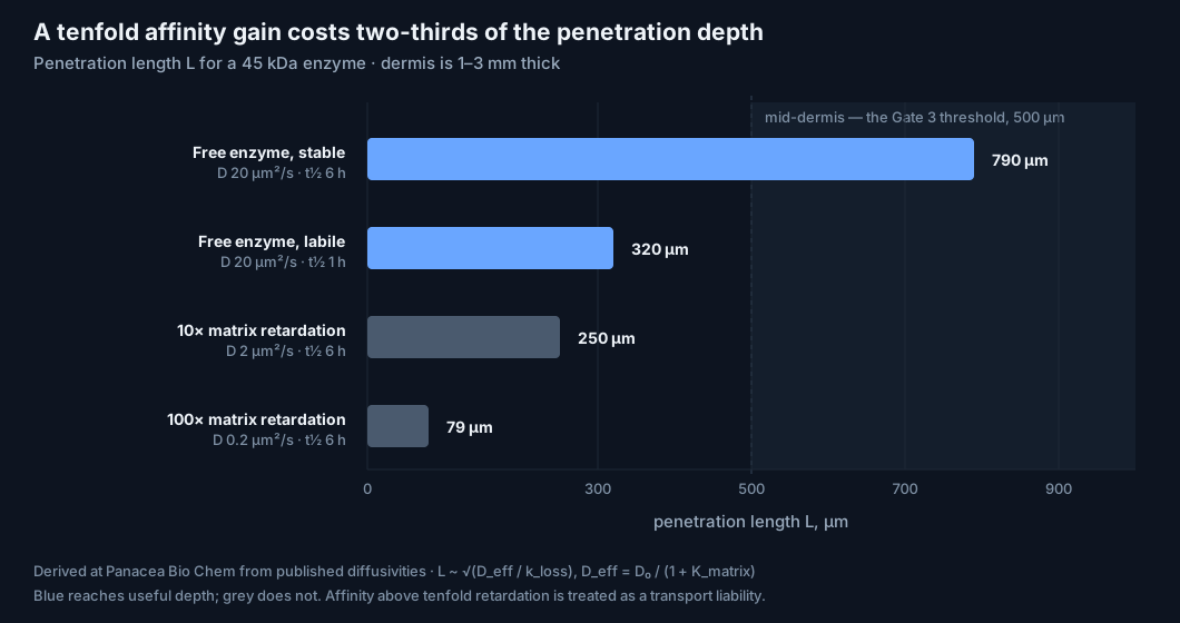
| Scenario | Deff (µm²/s) | Enzyme half-life | Penetration L | Reaches mid-dermis? |
|---|---|---|---|---|
| Free enzyme, stable | 20 | 6 h | 790 µm | Yes |
| Free enzyme, labile | 20 | 1 h | 320 µm | Partly |
| 10× matrix retardation | 2 | 6 h | 250 µm | No |
| 100× matrix retardation | 0.2 | 6 h | 79 µm | Surface only |
A tenfold affinity gain costs two-thirds of the penetration depth. The product of reached volume and local dwell time passes through a maximum at intermediate affinity, and every instinct in delivery engineering pushes past it.
This is the binding-site barrier, well characterised in antibody delivery, arriving in a new setting. The published histology was performed on 4 µm sections, a geometry in which L is irrelevant by construction. Nothing in that dataset tests transport at all.
The dose result
If 5 µM is the working concentration, the protein requirement is far smaller than intuition suggests.
| Target volume | Volume | Enzyme required (45 kDa) |
|---|---|---|
| 10 cm² forearm patch, 2 mm deep | 2 mL | 0.45 mg |
| Full facial dermis | ≈60 mL | 14 mg |
| Whole-body dermis (1.8 m², 2 mm) | 3.6 L | 810 mg |
Whole-body dermal coverage lands under one gram — inside the range at which therapeutic antibodies are routinely dosed. The enzyme is not the scarce resource. Access is.
What this means for our design
We start with free soluble enzyme and measure the depth profile of repair, not of fluorescent label. An enzyme can distribute to 500 µm while repairing only the outer 50 µm, and only a depth-resolved chemical readout distinguishes those two cases.
Matrix affinity is added only after a measured residence deficit, and any affinity above tenfold retardation is treated as a transport liability requiring explicit justification.
§8
Hypothesis 4 — bioelectric coupling
Hypothesis. Glycation damage degrades not only the mechanical matrix but the substrate on which cells read electrical and adhesive cues. Enzymatic repair should therefore be measurable as a restored response to stimulation, not only as a lower adduct count — and that response is a far more sensitive endpoint than stiffness.
The electrostatic argument, and the version of it that fails
CML formation converts a lysine ε-amine, protonated and carrying +1 at physiological pH, into a secondary amine bearing a carboxylate. Each modification event is a local charge swing of one to two units in the negative direction. Repair reverses it exactly.
The obvious extension is that bulk matrix fixed-charge density changes with age and is restored by the enzyme. That argument fails on arithmetic and we abandon it. At 1.4 mmol CML/mol Lys, only 0.14% of lysines are modified — roughly 40–80 µM of charge change against a glycosaminoglycan fixed-charge background one to three orders of magnitude larger. Bulk electrostatics do not move.
What survives is the site-specific version. Modification is strongly non-uniform, with some residues heavily hit and others untouched. A charge reversal on 0.14% of lysines is electrostatically trivial and biologically decisive if those lysines sit in integrin-binding or cell-adhesion motifs. The question is not how much charge changed. It is which residues changed.
What electrical stimulation actually does
Direct-current fields of physiological strength drive a large increase in small extracellular vesicle output. The mechanism is membrane electropolarisation recruiting lipid-raft and ceramide-rich domains, which drive inward budding into multivesicular bodies. Blocking ceramide synthesis or disrupting rafts collapses the effect to a few percent of stimulated output; blocking PI3K removes about half.
| Feature | Observation | Consequence for design |
|---|---|---|
| Dose window | Output rises to ≈200 mV/mm, then declines at 300 | Non-monotonic; more field is worse |
| Current type | Direct current effective, alternating much less so | Polarisation persistence is the driver |
| Vesicle class | Exosomes rise; shed microvesicles do not | Pathway-specific, not general membrane stress |
| Cargo | Benefit tracks yield, not activity per vesicle | More vesicles, not better vesicles |
| Persistence | Output falls rapidly once the field is withdrawn | No durable reprogramming from the field alone |
The dose window is worth dwelling on. The same stimulus that is regenerative at 200 mV/mm is less effective at 300. That is the identical structure as the peroxide question in §5 and the affinity optimum in §7: three independent variables in this system, each with an interior optimum, each of which the instinct of the field is to maximise.
The experiment that tests the coupling
Fibroblast galvanotaxis — directed migration in an applied field — requires integrin-mediated traction against the matrix. If CML on collagen degrades integrin engagement at specific sites, aged matrix should support a measurably weaker galvanotactic response than young matrix, and enzymatic repair should recover part of it.
| Arm | What it isolates |
|---|---|
| Untreated | Baseline response of aged matrix |
| Field only, 200 mV/mm DC | What bioelectric activation achieves on damaged substrate |
| Enzyme only | What chemical repair achieves without stimulation |
| Enzyme then field | Whether repair restores responsiveness to the cue |
The hypothesis predicts super-additivity in the fourth arm. If repair merely adds, the two mechanisms are independent and should be developed separately. If the fourth arm exceeds the sum, matrix damage was limiting the tissue's response to its own regenerative signalling — which would be a more important result than any adduct count.
Controls that are not optional
Electrode products — pH shift, electrolysis, metal ions — reproduce parts of the response and must be excluded with sham-electrode and media-transfer arms. Vesicle preparations must be characterised rather than assumed to be exosomes. And because the enzyme generates peroxide while the field activates redox-sensitive signalling, the peroxide-matched arm from §5 carries into this design unchanged.
On RAGE. The assumption that removing CML suppresses RAGE signalling is not safe. Purified protein carrying only CML has been reported not to bind or activate the receptor in some systems, while selected CML-bearing peptides and CML-modified albumin do bind under other conditions. Any inflammatory improvement we report must be shown to be RAGE-dependent by receptor blockade or knockout, never inferred from the adduct falling.
§9
Hypothesis 5 — enzyme space and cross-link scission
Hypothesis. CML repair is the easiest member of a family of reversals, not the hardest, and it addresses neither the most abundant nor the mechanically important lesion in aged matrix. The arithmetic says the field has optimised the wrong target first.
The abundance comparison nobody runs
| Lesion | Aged skin collagen | Molar concentration | Chemical class | Load-bearing? |
|---|---|---|---|---|
| CML | ≈1.4 mmol/mol Lys at 80 y | ≈40 µM | Side-chain adduct | No |
| Glucosepane | ≈2,000 pmol/mg collagen | ≈200 µM | Lys–Arg covalent cross-link | Yes |
Glucosepane outnumbers CML roughly five to one in aged dermis, rises to about 5,000 pmol/mg in diabetes, and exceeds every other known AGE cross-link in collagen and elastin by two to three orders of magnitude. It reaches roughly one cross-link per two to five collagen molecules and is estimated to reduce collagen digestibility 30- to 45-fold.
CML is the more tractable chemistry and the lesser lesion. A programme that removes CML and reports restored tissue mechanics has almost certainly measured something other than what it claims — which is why §14 requires glucosepane and pentosidine to be explicitly unchanged.
Why repair is the only route
Skin collagen has a half-life of about fifteen years. On that timescale, damage accumulating linearly with age is effectively permanent: the tissue cannot dilute it out. Glucosepane cross-linking specifically obstructs the matrix metalloproteinase step that normally initiates collagen breakdown, so the lesion blocks its own removal pathway. That is what distinguishes this class of damage from almost everything else in ageing biology, and it is why enzymatic reversal — rather than stimulation of turnover — is the correct intervention category.
Three deglycation mechanisms already exist in nature
| Mechanism | Representative chemistry | Substrate reached | Limit |
|---|---|---|---|
| Oxidative | FAD oxidases on fructosyl-amines → amine + glucosone + H₂O₂ | Free and low-MW glycated amines | Inert on protein-bound adducts |
| Phosphorylative | Fructosamine-3-kinase; fructoselysine-6-kinase | Fructosamine on proteins and free | Only Amadori products, which CML is not |
| Hydrolytic | Deglycase converting fructosamine-6-phosphate to glucose-6-phosphate + lysine | Phosphorylated adducts | Requires a prior kinase step; intracellular |
The oxidative family is the direct ancestor. Its structures are solved, it shares the monomeric sarcosine oxidase fold, and it has sat at the same barrier for three decades. The engineered CML enzyme is the first member of that family to cross it — and that achievement is a statement about access, not about chemistry.
The phosphorylative route deserves one line of elimination. It is attractive because it destabilises the adduct rather than oxidising it, and produces no peroxide. It is inapplicable here: it acts on Amadori products, and CML is a terminal oxidation product with no sugar left to phosphorylate. Mechanistic elegance does not transfer across chemical class.
A second fold is available, and we intend to take it
An N-alkylglycine oxidase isolated from a soil fungus in 1999 oxidised free CML to the corresponding amine, glyoxylate and peroxide — the same chemistry, 27 years early, on free substrate only. Its sequence was never reported and the isolate is unavailable.
Recovering that scaffold by re-isolation from environmental samples is a genuinely distinct engineering start point rather than a reproduction of existing work. A second fold performing this chemistry would immediately reveal which features are essential and which are historical accident — information no amount of further evolution on the current scaffold can produce.
Cross-link scission is a different problem, and it is tractable
Removing an adduct restores a side chain. Cleaving a cross-link must break a specific covalent bridge while leaving intact the enzymatic lysyl-oxidase-derived cross-links that give collagen its tensile strength. Selectivity, not reactivity, is the whole difficulty — an unselective cross-link breaker is a collagenase.
The area is further along than it appears. Enzymes capable of degrading peptide-bound glucosepane have been disclosed, with cleavage releasing citrulline from the lesion and no activity from the earlier small-molecule cross-link breaker compounds in the same assay. A separate 2026 chemical route reports a catalyst-trap/redox strategy for selective glucosepane cleavage, exploiting the fact that the aged collagen environment biases chemistry toward cross-link-bearing sites.
That last point is the interesting one, and it generalises. The lesion sits in a distinctive local environment — strained, sterically crowded, conformationally distinct from native collagen — and that environment is itself a targeting handle. The same logic applies to the inaccessible CML population identified in §7: sites resistant to an enzyme may be reachable by chemistry that the surrounding structure selects for.
§10
Programme architecture
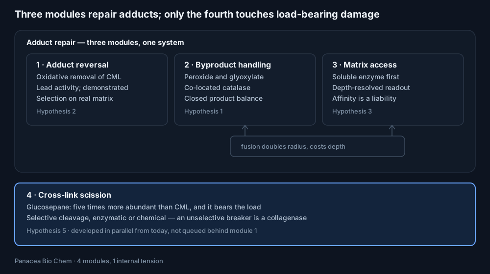
Modules 1 to 3 form one system because they share a route and a schedule. Module 4 does not, and queueing it behind the others would mean arriving at the mechanics question a decade late.
The tension drawn between modules 2 and 3 is the one architectural decision we have already taken: the tightest peroxide architecture (a fusion or conjugate) buys proximity by doubling hydrodynamic radius, which is paid for directly in penetration depth. Constraint 4 is traded against constraint 2, and constraint 2 ranks higher — so co-retention in a shared local phase is the default, and a fusion needs an explicit case.
Each module is characterised separately before any combination. The reason is diagnostic, not procedural: a combined system that underperforms gives no information about which module failed, and with four modules and three plausible failure modes each, the combinatorics defeat any reasonable experimental budget.
§11
The five-gate decision programme
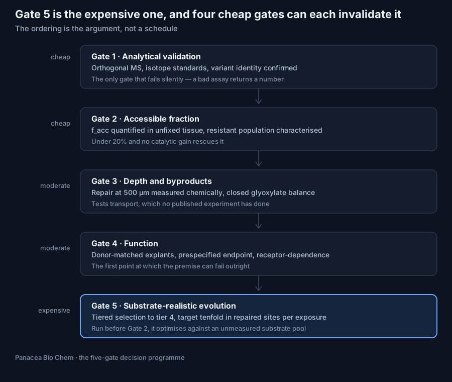
The ordering is the argument. Gates 1 to 4 are cheap relative to Gate 5, and each of them can invalidate it. A directed-evolution campaign launched before Gate 2 is optimising an enzyme against an unmeasured substrate pool; launched before Gate 4, it is optimising against an unproven target.
Gate 1 sits first because it is the only one whose failure is silent. Gate 2 can tell us the accessible fraction is under 20%, Gate 3 can tell us the enzyme never reaches mid-dermis, Gate 4 can tell us repair changes nothing biological — and all three of those arrive as results. An unvalidated assay does not return an error. It returns a number, and every subsequent decision inherits it.
What each gate decides
| Gate | Passes if | Fails if | Consequence of failure |
|---|---|---|---|
| 1 | Lysine restoration confirmed at mapped sites, orthogonal protease, variant identity established | Repair signal does not survive a non-antibody readout | Everything downstream is unsafe to interpret; stop and rebuild the analytics |
| 2 | facc quantified in unfixed tissue with the resistant population characterised | facc < 20% | Structural chemistry problem, not an enzymology problem; redirect to §9 |
| 3 | Chemical repair demonstrated at ≥ 500 µm; closed product balance, no oxalate rise | Repair confined to the outer tens of microns, or byproducts unaccounted | Delivery redesign before any further catalytic work |
| 4 | Prespecified cellular endpoint exceeds donor-matched variability, receptor-dependence shown | CML falls and nothing biological moves | CML is a marker, not a driver; value shifts to the platform (§15) |
| 5 | kcat/Km improved ≥ 10×, measured at tier 4 | Gains appear only at tier 1 | The screen is still selecting on the wrong substrate; return to §6 |
Gate 5 is deliberately last and deliberately expensive. It is the step everyone else starts with.
§12
Controls, confounders and the analytics that decide
Four measurement artefacts in this area are capable of producing a convincing, publishable, entirely false result. Each has a specific fix, and each is written into our protocols rather than left to judgement at analysis time.
The antibody problem
The published lens preparation gave 45% removal by mass spectrometry and 78% by ELISA on identical material. A supplementary ELISA returned 134 ± 61% repair at low initial modification.
Antibody readouts in this assay are not a noisy version of the truth. Near background they are uninterpretable, and anti-AGE antibodies preferentially recognise CML epitopes in ways that depend on surrounding structure — which is exactly what the enzyme changes.
Our rule. Immunohistochemistry is acceptable for spatial distribution and nothing else. Every quantitative claim requires LC–MS/MS with isotope-labelled internal standards.
The protease artefact
Restoring lysine restores a trypsin cleavage site. A repaired protein is therefore digested differently from its damaged counterpart, which changes peptide recovery, which changes apparent site occupancy — in the direction that flatters the result.
This artefact inflates measured repair without any repair occurring. Site-level comparisons must use an orthogonal protease such as Glu-C, or account explicitly for the shift in digestion pattern.
The endotoxin problem
The enzyme is bacterially expressed. Any inflammatory endpoint measured on a bacterially derived protein preparation is confounded by residual lipopolysaccharide unless purity, endotoxin load and aggregation state are measured on the actual material used.
Given that reduced inflammatory signalling is the most likely first benefit anyone will claim, this is the single most dangerous uncontrolled variable in the programme.
The variant problem
The published site-resolved proteomics were generated with an earlier evolved variant, with the inactive comparator from an earlier generation still. The deposited mass-spectrometry filenames carry those generation numbers.
Any work we do against that dataset must first establish which protein we actually hold, by intact-mass and peptide-mapping confirmation, before interpreting a single site. Two variants three evolutionary rounds apart are not the same molecule, and the headline kinetics and the site map do not describe the same protein.
The control matrix
No arm here is optional, and three of them are the experiment rather than its controls.
| Arm | What it isolates |
|---|---|
| Untreated tissue | Baseline |
| Catalytically inactive enzyme, matched purity | Effects of the protein preparation, not the chemistry |
| Active enzyme | Chemical repair |
| Catalase alone | Independent effect of peroxide disposal |
| Active enzyme + catalase | Whether peroxide removal helps or harms |
| Peroxide-matched, no enzyme | Whether oxidative signalling alone explains the response |
| Receptor-blocked or knockout | Whether inflammatory change is receptor-mediated |
| Unmodified substrate | Off-target oxidation of native residues |
Donor-matched tissue, independent biological replicates, randomisation and blinded analysis throughout. Donor-to-donor variation in AGE burden spans the entire effect size being sought; unmatched designs will not resolve it.
The full AGE profile
CML, carboxyethyl-lysine, pentosidine and glucosepane are measured before and after, in every tissue experiment.
If CML falls and the others do not move, that is the correct selective result and we report it as such. The failure mode to guard against is the opposite: a broad AGE decline, which would indicate non-specific oxidative degradation of the matrix rather than targeted repair.
§13
Predicted outcomes
The durability result
A single 60% clearance returns the CML burden of an 80-year-old's dermal collagen to that of a 33-year-old, and it takes roughly 47 years to come back.
This is the most consequential number in the document, and it follows from two linear relationships and nothing else. CML in human skin collagen accumulates at 0.018 mmol/mol Lys per year. Skin collagen has a half-life of about fifteen years, so the burden is not diluted by turnover — it integrates.
An 80-year-old therefore carries about 1.42 mmol CML/mol Lys. Remove 60%, which is at the low end of what has already been demonstrated on aged tissue, and 0.57 remains. Solve the accumulation line backwards:
| Removal achieved | CML remaining (mmol/mol Lys) | Equivalent collagen age | Years to re-accumulate |
|---|---|---|---|
| 40% | 0.85 | 48 years | 32 |
| 60% | 0.57 | 33 years | 47 |
| 70% | 0.43 | 25 years | 55 |
| 90% | 0.14 | 9 years | 71 |
The asymmetry is the whole point. Damage that takes a lifetime to form takes a lifetime to re-form. This is not a chronic therapy. One intervention per decade, or less, is sufficient to hold the burden down.
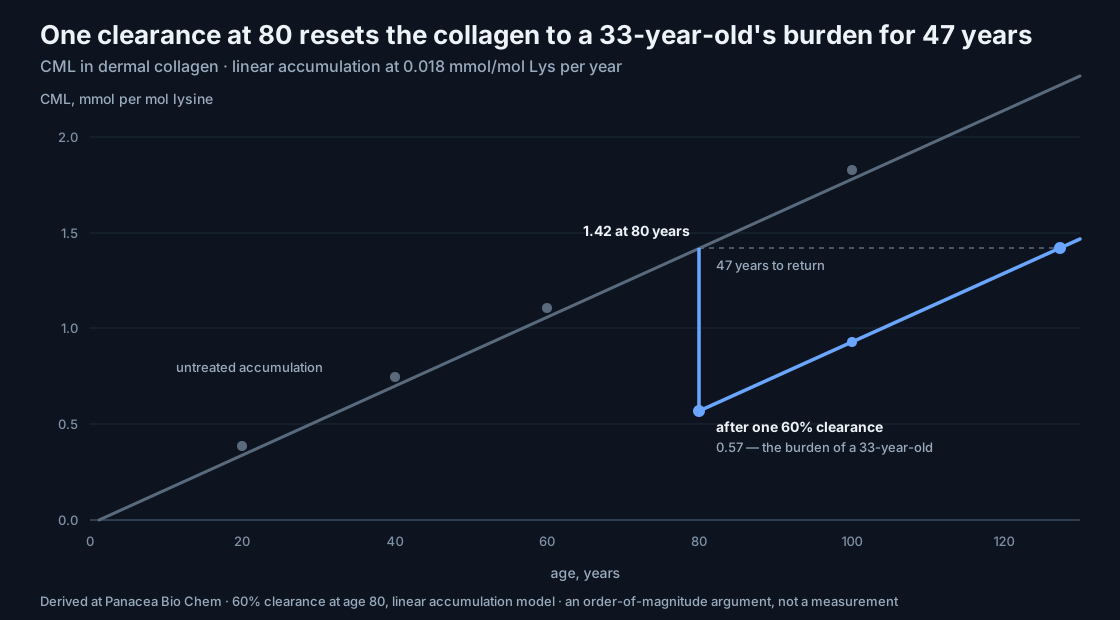
The slope is the same before and after. Nothing about the intervention changes the rate at which damage re-forms — which is exactly why the effect lasts.
That changes the economics and the risk profile together. It converts the immunogenicity problem from a recurring-dose liability into a tolerable one, and it makes a locally administered bacterial enzyme a far more reasonable proposition than it first appears.
The honest boundary on that claim: this is an adduct-burden equivalence, not a biological-age claim. It says the chemistry of the matrix is reset. Whether the tissue behaves younger is Gate 4, and nothing above answers it.
Three outcome classes
Class I — near-certain. Depth-resolved chemical repair in intact tissue. The chemistry is demonstrated and the transport model says a soluble 45 kDa enzyme reaches 300–800 µm. The quantity genuinely unknown is the accessible fraction in unfixed tissue, where the published 55–70% is an upper bound. We expect 20–50%, and we expect the resistant population to be sharply bimodal rather than a tail.
Class II — plausible. Restored cellular responsiveness: reduced pathological signalling, better fibroblast adhesion and migration, recovered galvanotactic response. This is more likely to appear than any change in bulk stiffness, because it depends on site-level recognition rather than on cross-link density. It is also the first endpoint at which the programme could fail outright, which is why we reach it as early as the gate structure allows.
Class III — the astonishing case. Super-additivity between matrix repair and bioelectric stimulation, meaning that accumulated chemical damage was limiting the tissue's response to its own regenerative signalling. If that holds, the implication is larger than the enzyme: a substantial part of what is measured as age-related loss of regenerative capacity would be not cellular decline but a degraded substrate, recoverable by chemistry rather than by cell replacement. Paired with selective cross-link scission, that is a mechanical and functional restoration rather than a biomarker correction.
§14
Thresholds
Internal decision criteria, set before any data exist, in the order they must be met. Setting them now is the point: a threshold written after the result is not a threshold.
| Measurement | First meaningful milestone | Gate |
|---|---|---|
| Chemical identity | Lysine restoration confirmed at mapped sites by orthogonal MS, with isotope standards | 1 |
| Accessible fraction | facc quantified in unfixed tissue, with the resistant population characterised | 2 |
| Penetration | Repair demonstrated at ≥ 500 µm depth, measured chemically, not by label | 3 |
| Byproducts | Closed product balance for glyoxylate and peroxide; no oxalate rise | 3 |
| Function | Prespecified cellular endpoint exceeding donor-matched variability, receptor-dependence shown | 4 |
| Cross-links | Glucosepane and pentosidine explicitly unchanged, confirming selectivity | 4 |
| Catalytic efficiency | kcat/Km improved ≥ 10× — not kcat alone | 5 |
The last row is the one most likely to be quietly relaxed under pressure, so it is stated twice. A tenfold kcat gain with unchanged kcat/Km does not meet this threshold and does not advance the programme, however good it looks in a figure.
§15
Failure modes worth naming
Three negative results are plausible enough to plan for. In each case we state now where the value goes, so that a bad result is a redirection rather than a sunk programme.
If CML falls by 60% and nothing biological moves. CML is a marker rather than a driver in that tissue. The programme's value shifts entirely to the platform — a demonstrated method for reversing a covalent protein modification in situ, retargetable at lesions that do drive pathology. That is still a substantial asset, and the modules in §10 are built to be retargeted rather than rebuilt.
If the accessible fraction in intact tissue proves to be under 20%. No amount of catalytic improvement rescues it. The problem becomes structural chemistry rather than enzymology, and the correct response is the environment-selective chemistry described at the end of §9, not another evolution campaign.
If repair succeeds and mechanics do not change. That is the predicted result, not a failure. CML is an adduct, not a bridge, and §9 says in advance that mechanics should not move. It is the strongest possible argument for funding cross-link scission, and we will treat it as one rather than as a disappointment.
The failure mode we do not plan for, because it cannot be recovered from, is a false positive at Gate 1. An unvalidated assay does not return an error. It returns a number, and every subsequent decision inherits it.
§16
Basis and provenance
This section separates what we take from the published record from what we derive here. The distinction is deliberate and load-bearing: the published values are the ground this programme is built on, and the derived figures are the programme's own argument. Readers should be able to tell which is which without asking.
Published values the programme rests on
| Figure used | Source |
|---|---|
| CML accumulation in skin collagen: 0.018 mmol/mol Lys per year (non-diabetic), 0.027 (diabetic) | Effects of age and diabetes on glycation of human skin collagen; age-dependent CML and carboxymethyl-hydroxylysine accumulation in skin collagen · Biochemistry, 1991 |
| Glucosepane ≈2,000 pmol/mg in aged skin collagen, ≈5,000 in diabetes; 100–1,000× other cross-links | Glucosepane reference summary |
| Glucosepane as the major senescent ECM cross-link; ≈500 pmol/mg in GBM; 30–45× reduction in collagen digestibility | J. Biol. Chem., glucosepane in senescent human ECM |
| Skin collagen half-life ≈15 years; glucosepane obstructs MMP-mediated breakdown | Reviews of glucosepane clearance approaches |
| Enzymatic cleavage of peptide-bound glucosepane, releasing citrulline; no activity from earlier small-molecule breakers | WO2020215043A1, AGE-breaking biocatalysts |
| Chemical catalyst-trap/redox route to selective glucosepane cleavage, 2026 | ChemRxiv preprint |
| Preferential intramolecular glucosepane sites in type I collagen; effect on tensile and lateral moduli | Thermodynamic and mechanical-properties studies |
| Fructosamine oxidase structure; flexible active-site loops as determinants of substrate size | Crystal structure of fructosamine oxidase; Amadoriase I structures |
| Three distinct deglycation mechanisms: oxidase, kinase, hydrolytic deglycase | Enzymatic deglycation overview; fructosyl-amino acid oxidases; EC 1.5.3.25 |
| Catalase turnover up to 2.8 × 10⁶ s⁻¹ | Catalase turnover number and catalytic efficiency |
| Electric fields drive ceramide-dependent sEV budding; 200 mV/mm optimum, decline at 300; DC over AC; yield not per-vesicle activity | Commun Biol, 2 July 2026 |
| Electrical stimulation modulates sEV secretion and cargo; nSMase2 dependence | Tissue Eng Regen Med, 15 July 2026; EV production engineering reviews |
| Macromolecule diffusivity in dermis 10–20 µm²/s; dermis 1–3 mm thick | Diffusion kinetics in human skin; dermal interstitial fluid reviews |
| Binding-site barrier; interstitial diffusion coefficient ≈1.3 × 10⁻⁷ cm²/s; affinity–penetration trade-off | Antibody penetration modelling; antibody fragments and tumour penetration |
| ECM composition governs macromolecule diffusion; collagen the dominant barrier | IgG diffusion and ECM composition studies |
| CML present in epidermis; anti-AGE antibodies preferentially recognise CML epitopes | CML in human epidermis; quantitative CML model |
| N-alkylglycine oxidase oxidising free CML, 1999 | Soil fungus isolate; sequence never reported |
| Kinetic constants, mutation series, tissue staining percentages, site-resolved proteomics, peroxide output | The primary 2026 publication, its supplementary tables, its transparent peer-review file and the deposited proteomics dataset |
Figures derived at Panacea Bio Chem
The following are calculated in this document from the sourced values above. They are not literature measurements, and each rests on stated assumptions about dermal collagen density, lysine content and enzyme mass. Each is an order-of-magnitude argument rather than a measurement.
- The ≈40 µM matrix CML concentration
- The sub-saturating regime and the pseudo-first-order 3.9-hour half-time
- The predicted 96% removal, the resulting 26–41 point shortfall, and the two-population substrate argument
- The accessible-fraction estimate and its upper-bound interpretation
- Penetration lengths and the affinity–depth trade-off table
- The enzyme dose table
- The age-equivalence and re-accumulation table
- The catalase rate-mismatch arithmetic and the coupled net reaction
- The P(NAC) formulation and the tiered selection ladder
- The seven-constraint ranking, the five gates, the thresholds and the control matrix
Two sources needing confirmation. The skin collagen regression and the glucosepane abundance paper returned indexed page content but refused direct retrieval. Their figures should be confirmed against the published articles before being used quantitatively in any external filing.
Citation
How to cite this paper
Dicoias, B. (2026). Enzymatic Reversal of Advanced Glycation Damage: architecture, hypotheses and the five-gate decision programme. Panacea Bio Chem Ltd. Published 31 October 2026. https://cmlase.org/programme/
@techreport{dicoias2026cmlase,
  author      = {Dicoias, Bogdan},
  title       = {Enzymatic Reversal of Advanced Glycation Damage:
                 architecture, hypotheses and the five-gate decision programme},
  institution = {Panacea Bio Chem Ltd},
  year        = {2026},
  month       = {10},
  type        = {Programme paper},
  url         = {https://cmlase.org/programme/}
}
A DOI is being registered for this paper; it will be added here and to the page metadata on deposit.
Scientific position
This is an independent research programme of Panacea Bio Chem Ltd, led by Bogdan Dicoias. The original CrGO-897/CMLase enzyme was reported by external researchers; Panacea Bio Chem does not claim authorship or ownership of that discovery, and this paper is an independent research direction arising from it.
CMLase remains experimental and is not an approved medical treatment. Nothing on this page is medical advice. See the scientific disclaimer.